News
Endoscope Working Principle
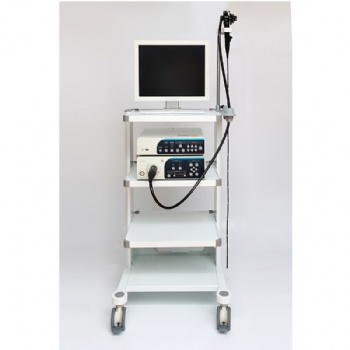
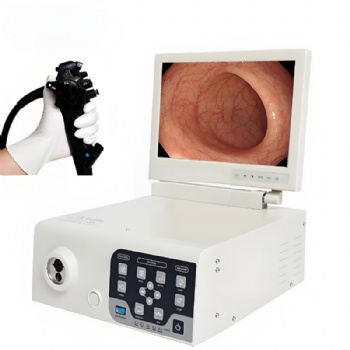
The endoscope is widely used in minimally invasive surgery clinical use as a low-light, high-resolution color digital electronic camera. By using a light-sensitive chip to achieve unprecedented high resolution and provide true reproduction of image color. This makes it easier to observe the patient's microscopic lesions, and its high-quality light source and low-loss digital magnification of the image can identify very small image details. It is very easy to operate and can be used for both diagnostic and surgical treatment procedures.
Working principle of the endoscope
It is a diagnostic method of gross morphology, using a high-resolution color camera to magnify the mucosa of the lesion, and through camera image processing, thus observing its surface morphology and changes in the terminal vascular network, and observing tiny lesions that are not visible to the naked eye.
Functional requirements
Camera integration full field of view clear, no blurring at the edge of the field of view. Signal output, to meet the clinical requirements. Camera with an automatic white balance function, by pressing the white balance key can automatically establish the color balance. The instrument toggle switch, and regulator should be operated flexibly and reliably, with no poor contact and misoperation. The connection of the various parts of the instrument should be reliable, no poor contact, loose or even off the electrical connectors and other phenomena. The connection between the interface and the endoscope should be easy to operate, correctly and reliably positioned, stable after locking, light area control technology, and very fine endoscope imaging quality as perfect.
 Send Email