News
Development of Medical Endoscope Camera System
The medical endoscope video camera system is suitable for all departments, and is widely used in otolaryngology, urology, gynecology, ophthalmology, orthopedics, neurosurgery, medical aesthetics and other departments in clinical practice. Laparoscopy, hysteroscopy, arthroscopy, foramenoscopy, fiberoptic bronchoscopy and other endoscopy fields. With the continuous development of medical technology, medical endoscope technology is also constantly improving. Let's take a look at the development process of medical endoscope video camera system and learn more about medical endoscope video camera system.
1. Development history of medical endoscope video camera system
(1) Rigid endoscope stage (1806--1932)
The rigid endoscope stage was pioneered by the Germans and consisted of a vase-shaped light source, candles and a series of lenses, mainly used for bladder and urethral examinations. The rigid endoscope developed in 1895 consists of three tubes arranged in concentric circles. The gastroscope was improved in 1911, but the inability to observe after the lens became dirty was the main defect, but the gastroscope was still in the lead until 1932.
(2) Semi-flexible endoscope stage (1932-1957)
Schindler developed gastroscopes in cooperation with excellent instrument operators in 1928, and finally succeeded in 1932, named Wolf-Schinder gastroscopes. After that, everyone modified it to make it more functional and practical.
(3) Fiber optic endoscopy stage (1957 to present)
In 1954, the British Hopkins and Kapany invented the optical fiber technology. In 1957, Hirschowitz and his assistants demonstrated their self-developed fiberoptic endoscope at the American Society of Gastroscope. In the early 1960s, Japan adopted an external cold light source, which greatly increased the brightness and further expanded the field of vision. In the past 10 years, with the continuous improvement of accessory devices, fiber endoscopes can be used not only for diagnosis, but also for surgical treatment.
(4) The era of electronic endoscopy (after 1983)
In 1983, the electronic camera endoscope was successfully developed. The front end of the mirror is equipped with a high-sensitivity miniature camera. The recorded images are transmitted to the television information processing system in the form of electrical signals, and then the signals are converted into images that can be seen on the television monitor.
2. The composition of the medical endoscope video camera system
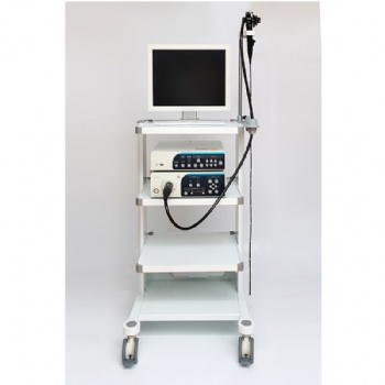
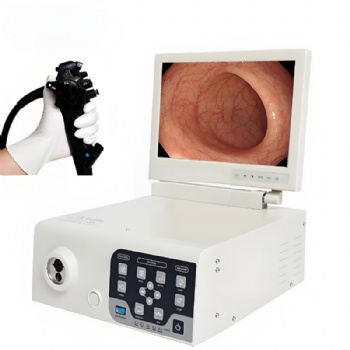
The medical endoscope video camera system consists of an endoscope optical interface, a camera head and a camera system. The camera system consists of a camera, a power cord and various connecting wires; the current camera includes a single-chip camera and a three-chip camera; the optical interface of the endoscope is suitable for: cystoscope, laparoscope, sinusoscope, support laryngoscope, otoscope, uterine endoscopes, etc.
3. The role of medical endoscope video camera system
(1) Guide light, direct light from a strong light source outside into the organ to illuminate the inspection site.
(2) Guide image, transmit the image reflecting the endoscopic situation of the organ, through the monitor, it is convenient for the doctor to observe the clear and detailed intracavity tissue, and the endoscope video camera provides the guarantee for the doctor to operate safely and delicately.
Categories
Contact Us
- +86-18018467613
- +86-13357930108
- info@82tech.com


